

Please use this identifier to cite or link to this item:
https://hdl.handle.net/10216/116257Full metadata record
| DC Field | Value | Language |
|---|---|---|
| dc.creator | Yu Wang | |
| dc.creator | Yating Chen | |
| dc.creator | Ningning Yan | |
| dc.creator | Longfei Zheng | |
| dc.creator | Nilanjan Dey | |
| dc.creator | Amira S. Ashour | |
| dc.creator | V. Rajinikanth | |
| dc.creator | João Manuel R. S. Tavares | |
| dc.creator | Fuqian Shi | |
| dc.date.accessioned | 2022-09-08T18:44:38Z | - |
| dc.date.available | 2022-09-08T18:44:38Z | - |
| dc.date.issued | 2019-01 | |
| dc.identifier.issn | 1568-4946 | |
| dc.identifier.other | sigarra:293683 | |
| dc.identifier.uri | https://hdl.handle.net/10216/116257 | - |
| dc.description.abstract | Hepatic granuloma develops in the early stage of liver cirrhosis which can seriously injury liver health. At present, the assessment of medical microscopic images is necessary for various diseases and the exploiting of artificial intelligence technology to assist pathology doctors in pre-diagnosis is the trend of future medical development. In this article, we try to classify mice liver microscopic images of normal, granuloma-fibrosis 1 and granuloma-fibrosis2, using convolutional neural networks (CNNs) and two conventional machine learning methods: support vector machine (SVM) and random forest (RF). On account of the included small dataset of 30 mice liver microscopic images, the proposed work included a preprocessing stage to deal with the problem of insufficient image number, which included the cropping of the original microscopic images to small patches, and the disorderly recombination after cropping and labeling the cropped patches In addition, recognizable texture features are extracted and selected using gray the level co-occurrence matrix (GLCM), local binary pattern (LBP) and Pearson correlation coefficient (PCC), respectively. The results established a classification accuracy of 82.78% of the proposed CNN based classifiers to classify 3 types of images. In addition, the confusion matrix figures out that the accuracy of the classification results using the proposed CNNs based classifiers for the normal class, granuloma-fibrosisl, and granuloma-fibrosis2 were 92.5%, 76.67%, and 79.17%, respectively. The comparative study of the proposed CNN based classifier and the SVM and RF proved the superiority of the CNNs showing its promising performance for clinical cases. | |
| dc.language.iso | eng | |
| dc.rights | openAccess | |
| dc.subject | Ciências da Saúde, Ciências Tecnológicas | |
| dc.subject | Health sciences, Technological sciences | |
| dc.title | Classification of mice hepatic granuloma microscopic images based on a deep convolutional neural network | |
| dc.type | Artigo em Revista Científica Internacional | |
| dc.contributor.uporto | Faculdade de Engenharia | |
| dc.identifier.doi | 10.1016/j.asoc.2018.10.006 | |
| Appears in Collections: | FEUP - Artigo em Revista Científica Internacional | |
Files in This Item:
| File | Description | Size | Format | |
|---|---|---|---|---|
| 293683.png | 1st Page | 178.79 kB | image/png | 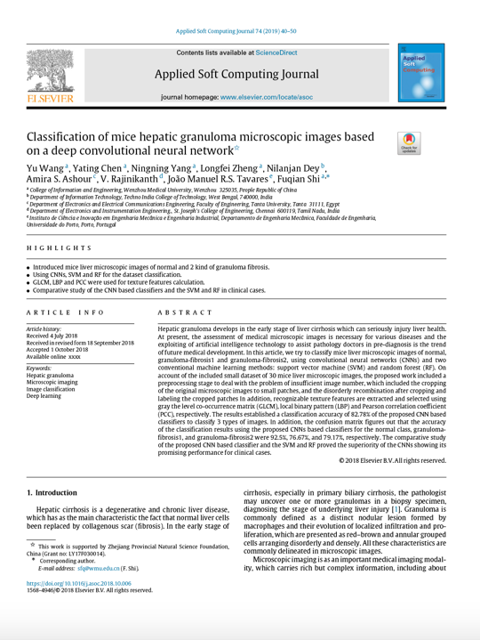 View/Open |
| 293683.1.pdf | Paper draft | 1.15 MB | Adobe PDF |  View/Open |
Items in DSpace are protected by copyright, with all rights reserved, unless otherwise indicated.